9/06/2011

Cardiac Arrhythmias 1/3 - Heart Physiology - USMLE Step 1

Cardiac Arrhythmias 1/3 - Heart Physiology - USMLE Step 1


▶▶▶ Watch More Videos at www.DrNajeebLectures.com ◀◀◀
Video Rating: 5 / 5


Orignal From: Cardiac Arrhythmias 1/3 - Heart Physiology - USMLE Step 1

Adrenergic System and Ventricular Arrhythmias in Myocardial Infarction Reviews

Adrenergic System and Ventricular Arrhythmias in Myocardial Infarction


ventricular system - click on the image below for more information.


ventricular system


This book presents new aspects on electrophysiological mechanisms and catecholaminergic contributions in the setting of acute and chronic myocardial ischemia. Special emphasis is placed on the full scope from basic molecular and cellular mechanisms to experimental models of close clinical proximity. A number of internationally distinguished scientists present their latest findings in this significant research area within the perimeter of cardiovascular disease which continues to lead mortality s


Adrenergic System and Ventricular Arrhythmias in Myocardial Infarction


Click on the button for more ventricular system information and reviews.


Betting Systems - Great Gambling Systems.
Gambling Systems And Strategy Tips For Online Sports Betting, Internet Horse Racing & Casino. New Arbitrage Betting Software.
Betting Systems - Great Gambling Systems.
Ventricular Fibrillation

Article by Michael A. Morales


Ventricular Fibrillation (also called v-fib or vf) is a lethal arrhythmia that originates in the ventricles. It commonly occurs in cardiac arrest patients and is the primary rhythm that AED's (automated external defibrillators) are looking for to initiate a shock to the victim of cardiac arrest

In v-fib there is no organized depolarization of the ventricles. The heart muscle simply quivers with no contraction to pump the blood. As a result, there is no pulse. There are a number of reasons that ventricular fibrillation can occur:

Electrolyte ImbalanceAcute Coronary SyndromesHeart failure DysrhythmiasHypertrophyIncreased sympathetic Nervous System Activity

The list really only provides a snap shot and is not all inclusive. The only known effective treatment for ventricular fibrillation is defibrillation. When defibrillation occurs, the heart is stopped for an instant and aloud to reset. You can think of it as kind of like rebooting your computer. Once reset, the heart may go back to an organized rhythm and blood flow can resume to the heart and vital organs.

Generally, CPR is performed while and AED is being accessed. CPR does not change ventricular fibrillation to a normal heart rhythm. CPR pumps the heart so that oxygen rich blood can continue to flow to the heart and vital organs until defibrillation is available. CPR buys the victims some time and may keep brain damage from occurring. Together early CPR and defibrillation saves lives if provided immediately and is currently the best defense against ventricular fibrillation for the victim of cardiac arrest.

Although the heart can go into a number of different rhythms in a cardiac arrest, ventricular fibrillation is one of the most common, and is treatable if address quickly. Without early defibrillation the hearts electrical system will cease to function. No electrical activity in the heart is a condition known as asystole or flat line. Defibrillation will correct asystole.

The 2005 American Heart Association guidelines recommend defibrillation within the first 3-5 minutes of a cardiac arrest. It is during this time period that the victim is more likely to be in v-fib and benefit from defibrillation.

Michael Morales


About the Author

Michael Morales is an EMT paramedic and director of education for Vital Ethics Inc., providing basic and advanced life support training and certification programs.

http://www.aclsclass.info/certification1.html

http://www.aclsclass.info/certification2.html

http://www.aclsclass.info/certification3.html


Orignal From: Adrenergic System and Ventricular Arrhythmias in Myocardial Infarction Reviews

Can food trigger Atrial Fibrillation?


Can food trigger Atrial Fibrillation?
Yes. I experience all your symptoms. I have atrial fibrillation with episodes lasting from 3 - 10 hours and am taking warfarin and beta blockers. I also have a hiatus hernia (without relux, just gas). Many of my fibrillations start immediately after a heavy meal or eating too quickly. Also if I lean forward and press on the diaphragm immediately after eating. I can't get the doctor to agree the connection - in fact he thinks I am up the creek, but I am convinced that they are connected. The hospital heart doctor also said that he had never heard of a connection. However my osteopath/acupuncturist actually took the connection for granted and says it is because the vagus (or vagal) nerve goes past the diaphragm and close to the heart and when the hernia is bad it triggers off the fibrillations. I asked my doctor to treat the hernia just to see what happens. He has put me on very strong digestive tablets and the hernia has subsided. I am waiting to see what happens if it flares up again. I hope no more fibs.
I would suggest that you check out the possibilty that you have a hiatus hernia remembering that you don't necessarily have to have reflux to prove you have one, just bad indigestion. Eat slowly and don't bend over after meals or even do very much for about half an hour and try not to squash the diaphragm.
I have now had acupuncture and can't believe how much more comfortable I am now after meals and no fibrillations for the last 2 1/2 weks . It is difficult to tell because I have recently been in hospital for a few days because the fibrillations got very frequent so the fibs may have stopped because of the tablets I was given and the injections of warfarin and because I had to drink potassium which controls the electrical activity in the heart. My reading was 2 instead of 5 and this was causing the fibs every other day. They gave me potassium tablets to take until I reach level 5 and I hope that my test on Wednesday reaches normal and I can stop taking them.

No real answers but would suggest that you check out the possibilty that you have a hiatus hernia remembering that you don't necessarily have to have reflux to prove you have one. Eat slowly, have small meals frequently instead of one big one and don't bend over after meals or even do very much for about half an hour and try not to squash the diaphragm. I too was frightened to eat and got quite paranoid but that feeling has now gone although I am very careful in what I eat now. No fried, spicy or fatty foods.
Do have your potassium levels checked (through a simple blood test) if you have not already done so because the level is very important. Mine was depleted through diuretics but many things can deplete it and it definitely triggers of electrical activity in the heart.The hospital coudn't wait to get the potassium into me and my warfarin levels up.

Good luck. Let me know how you get on and I will post anything I find through my search for the link that I firmly believe is there.
Patsy72Can food trigger A-fib? Certain food groups can increase the odds of creating a-fib in those with a history of preexisting a-fib or those with existing heart disease. Not to get too detailed Xanthines (found in chocolate) and caffeine (found in coffee and tea) are two common sources that have been implicated-- but what t you describe is not food induced a-fib but rather the food induced retching that triggered the a-fib . For the most part a-fib can be stabilized and treated medically -- What is really of concern in this whole matter is your protracted history of nausea and,bloating after eating or drinking anything- your Doctor should be actively searching for reasons why you have these symptoms ie hiatal hernia,gall bladder, ulcer,pancreas etc...Very good advice here about getting your "hiatal hernia" risk assessed. Often GI reflux can irritate your heart as well. I would recommend a consult to a gastroenterologist. Also keep in mind that whenever food or drink enters the stomach, blood flow is diverted to that area to assist with digestion. That means blood flow is diverted away from your heart (not alot). Have you had complete cardiac workup to determine if you have any coronary blockages. Even small diversions of blood away from the coronaries in the presence of coronary occlusions can trigger cardiac arrythmias and/or chest pain. Some people are very sensitive to this. My experience with recovering heart attack patients is to insist on rest for at least 30-45 minutes after meals to avoid any additional stress to the heart while food is being digested. Sitting upright for 30 minutes after meals also helps if you have hiatal hernia and/or reflux disease. Just some tips, hope it helps.

Orignal From: Can food trigger Atrial Fibrillation?

when reading the EKG of someone with right ventricular hypertrophy and right bundle branch block on the V1?


when reading the EKG of someone with right ventricular hypertrophy and right bundle branch block on the V1?
T wave inversion in V1 to V3 could be due to right bundle branch block or previous anterior wall myocardial infarction. T-wave inversion (what shows up in the three "abnormal" leads) can be caused by anything from hyperventilation to potassium depletion to positioning of the leads. It can, of course, also signify some non-specific heart disease, but in the absence of symptoms this is extremely difficult to know. I would move forward with a stress echo...t inversion in v1 alone is a normal phenomenon, if it extends to v2 and v3 it indicates ischemia.

Orignal From: when reading the EKG of someone with right ventricular hypertrophy and right bundle branch block on the V1?

Are Common Heart Skip Palpitations Dangerous?: Premature Ventricular and Atrial Contractions (PVCs and PACs)

Are Common Heart Skip Palpitations Dangerous?: Premature Ventricular and Atrial Contractions (PVCs and PACs)


pvc ventricular - click on the image below for more information.


pvc ventricular


INTRODUCTION: Skipped heartbeats occur in most, if not 100% of the general population, at some point during their lives. Some medical sources state that about half of us experience them on a relatively frequent basis. For some people however, these heart palpitations called "Premature Ventricular Contractions" and "Premature Atrial Contractions" (PVCs and PACs), occur at a frequency or forcefulness, that can be concerning to them and that can result in chronic anxiety and/or panic att


Are Common Heart Skip Palpitations Dangerous?: Premature Ventricular and Atrial Contractions (PVCs and PACs)


Click on the button for more pvc ventricular information and reviews.


Rocket Speed Training On A Budget 2.
A Speed & Agility Training Resource Guide on how to create your own speed & agility training aids, primarily out of Pvc pipe, which helps you save money & can give you more training options than traditional speed/agility tools.
Rocket Speed Training On A Budget 2.
Photoplethysmograph

Article by jekky


(721, 'Sites for measuring PPG br While pulse oximeters are a commonly used medical device the PPG derived from them is rarely displayed and is nominally only processed to determine heart rate PPGs can be obtained from transmissive absorption as at the finger tip or reflective as on the forehead br In outpatient setting pulse oximeters are commonly worn on the finger and ear However in cases of shock hypothermia etc blood flow to the periphery can be reduced resulting in a PPG without a discernible cardiac pulse In this case a PPG can be obtained from a pulse oximeter on the head with the most common sites being the ear nasal septum and forehead br PPGs can also be obtained from the vagina and esophagus br Uses br Premature Ventricular Contraction PVC can be seen in the PPG just as in the EKG and the Blood Pressure BP br Venous pulsations can be clearly seen in this PPG br Monitoring Heart Rate and Cardiac Cycle br Because the skin is so richly perfused it is relatively easy to detect the pulsatile component of the cardiac cycle The DC component of the signal is attributable to the bulk absorption of the skin tissue while the AC component is directly attributable to variation in blood volume in the skin caused by the pressure pulse of the cardiac cycle br The height of AC component of the photoplethysmogram is proportional to the pulse pressure the difference between the systolic and diastolic pressure in the arteries As seen in the figure showing Premature Ventricular Contractions PVCs the PPG pulse for the cardiac cycle with the PVC results in lower amplitude blood pressure and a PPG Ventricular Tachycardia and Ventricular Fibrillation can also be detected br Monitoring Respiration br The effects of Sodium Nitroprusside Nipride a peripheral vasodilator on the finger PPG of a sedated subject As expected the PPG amplitude increases after infusion and additionally the Respiratory Induced Variation RIV becomes enhanced br Respiration affects the cardiac cycle by varying the intrapleural pressure the pressure between the thoracic wall and the lungs Since the heart resides in the thoracic cavity between the lungs the partial pressure of inhaling and exhaling greatly influence the pressure on the vena cava and the filling of the right atrium This effect is often referred to as normal sinus arrhythmia br During inspiration intrapleural pressure decreases by up to 4 mm Hg which distends the right atrium allowing for faster filling from the vena cava increasing ventricular preload and increasing the stroke volume Conversely during expiration the heart is compressed decreasing cardiac efficiency and reducing stroke volume However the overall net effect of respiration is to act as pump for the cardiovascular system When the frequency and depth of respiration increases the venous return increases leading to increased cardiac output Shelley et al 2006 br Monitoring Depth of Anesthesia br Effects of an incision on a subject under general anesthesia on the photoplethysmograph PPG and blood pressure BP br Anesthesiologist must often judge subjectively whether a patient is sufficiently anesthetized for surgery As seen in the figure if a patient is not sufficiently anesthetized the sympathetic nervous system response to an incision can generate an immediate response in the amplitude of the PPG br Monitoring Hypo and Hyper volemia br Shamir Eidelman et al studied the interaction between inspiration and removal of 10 of a patient blood volume for blood banking before surgery Shamir Eidelman et al 1999 They found that blood loss could be detected both from the photoplethysmogram from a pulse oximeter and an arterial catheter Patients showed a decrease in the cardiac pulse amplitude caused by reduced cardiac preload during exhalation when the heart is being compressed br References br M Shamir L A Eidelman Y Floman L Kaplan and R Pi zov Pulse Oximetry Plethysmographic Waveform During Changes in Blood Volume Br J Anaesth vol 82 pp 178 181 1999 br K Shelley and S Shelley Pulse Oximeter Waveform Photoelectric Plethysmography in Clinical Monitoring Carol Lake R Hines and C Blitt Eds W B Saunders Company 2001 pp 420 428 br K H Shelley D H Jablonka A A Awad R G Stout H Rezkanna and D G Silverman What Is the Best Site for Measuring the Effect of Ventilation on the Pulse Oximeter Waveform Anesth Analg vol 103 pp 372 377 2006 br A T Reisner P A Shaltis D McCombie and H H Asada Utility of the Photoplethysmogram in Circulatory Monitoring Anesthesiology vol 108 pp 950 958 2008 br External links br A student project building a device for collecting PPGs br See also br Hemodynamics br Categories Cardiology Medical tests')


About the Author

I am China Crafts Suppliers writer, reports some information about bostitch air, cordless framing nailer.


Orignal From: Are Common Heart Skip Palpitations Dangerous?: Premature Ventricular and Atrial Contractions (PVCs and PACs)

9/01/2011

left ventricular systolic pressure and aoritic systolic pressure?


left ventricular systolic pressure and aoritic systolic pressure?
Aortic valve stenosis.

Coarctation proximal to the aortic measurement site.

Orignal From: left ventricular systolic pressure and aoritic systolic pressure?

Cardiac Resynchronization Therapy and Implantable Cardiac Defibrillators in Left Ventricular Systolic Dysfunction (Evidence Report/Technology Assessment)

Cardiac Resynchronization Therapy and Implantable Cardiac Defibrillators in Left Ventricular Systolic Dysfunction (Evidence Report/Technology Assessment)


ventricular defibrillator - click on the image below for more information.


ventricular defibrillator


Cardiac Resynchronization Therapy and Implantable Cardiac Defibrillators in Left Ventricular Systolic Dysfunction (Evidence Report/Technology Assessment)


Click on the button for more ventricular defibrillator information and reviews.


Orignal From: Cardiac Resynchronization Therapy and Implantable Cardiac Defibrillators in Left Ventricular Systolic Dysfunction (Evidence Report/Technology Assessment)

8/31/2011

BES FIB-EZXL3000 -inchThe Extreme Light-inch Rechargable LED Work Light

BES FIB-EZXL3000 -inchThe Extreme Light-inch Rechargable LED Work Light


v fib - click on the image below for more information.


  • Cordless, rechargeable, LED work light
  • 30 super bright LEDs
  • Swivel hook for easy hands free use
  • Includes charger

v fib


The BES cordless and rechargeable FIB-EZXL3000 "The Extreme Light" work light features 30 super bright LEDs and a swivel hook for easy hands free use. Includes charger.


BES FIB-EZXL3000 -inchThe Extreme Light-inch Rechargable LED Work Light


Click on the button for more v fib information and reviews.


V-Ecommerce
V-Ecommerce
V-Ecommerce
How To Use An Automated External Defibrillator (AED)

Article by Marcus Pierre


An automated external defibrillator (AED) is a device the size of a laptop that is used for medical emergencies, specifically cardiac arrest or when the heart stops beating. Sometimes, when a victim is in cardiac arrest, their heart start experiencing an irregular quivering caused by chaotic electrical activity in the heart cells.

This quivering is called ventricular fibrillation or V Fib for short. V Fib is treated by shocking the heart with controlled electric shock. This works because the heart muscle contains electrical cells. The AED is used to shock the heart that is in V Fib to a normal heart rhythm.

Studies show that if an AED is used within 3-5 minutes of cardiac arrest, the victim's chance of survival increases by approximately 70%. So you want to use the AED as soon as it becomes available.

Using the AED:

Once you get the AED at the vicitm's side -

1: Turn on the AED. Do this by wither pressing the on/off button or lifting the lid of the unit.

2: Follow the prompts exactly as directed. The device will instruct you to place the electrode pads on the bare skin of the victim. One of the pads are to be placed on the right upper chest of the victim, while the second pad is to be placed just under the left chest.

3: Once the pads are placed on the bare chese of the victim, you may be required to insert the pad connector to the AED. Some AEDs already have the pads preconnected to them.

4: Next you must make sure that no one (including yourself) is touching the victim. The machine will prompt that it is "analyzing." During the analysis, all CPR efforts must be paused in order for the AED to evaluate if the victim's heart is experiencing V Fib and can therefore be treated with a shock. This is one of the 2 time you must "clear" the victim of all bystander contact.

5: If the AED prompts that a shock is indicated, then you must "clear" the victim a second time ensuring that you or no bystanders are not in contact with the victim. If someone is touching the victim while the AED is delivering the shock, the bystander can experience a shock as well which can even render them unconscious. Once you see that victim is "clear," you must then press the "shock" button to deliver a shock. Fully automated AEDs will deliver the shock automatically.

6: Immediately after administering the shock, the defibrillator will instruct you to resume CPR. You must not remove the pads or turn off the unit at this time. Simply leave the pads on the victim's chest, leave the AED on and resume CPR beginning with chest compressions.Some of the new AED models have the CPR coaching feature which guides the rescuer in the CPR process.

AEDs are very simple to use as long as the instructions are followed completely. The information in this article does not substitute a CPR/AED course in which training is performed in its entirety. This information is provided solely as a reference to supplement CPR/AED training.

For more information or questions on AEDs please visit http://www.lifesaversny.com


About the Author

Marcus Pierre is a respiratory therapist, CPR instructor and AED consultant. He has worked at several hospitals in the New York area and has also trained many participants in CPR/AED & First Aid. He has serviced Regal Cinemas, the Governor's office, the Attorney General's office, DMV offices and many more NY state and personal clients. He is also the president of Lifesavers, Inc - a CPR & First Aid training company and an AED distributor. He can be contacted at http://www.lifesaversny.com


Orignal From: BES FIB-EZXL3000 -inchThe Extreme Light-inch Rechargable LED Work Light

What is 2nd degree AV block Mobitz type 1 Wenckebach?


What is 2nd degree AV block Mobitz type 1 Wenckebach?
AV block can be manifested as several different types. All of them being a conduction abnormality in your AV node. Try searching for just "Wenckebach" or "Mobitz type 1."

Ventricular bigeminy means that you have premature beats originating in the ventricles. The bigeminy part indicates that it was every other beat.

Orignal From: What is 2nd degree AV block Mobitz type 1 Wenckebach?

What is the difference between the thickness of the aorta and ventricular walls?


What is the difference between the thickness of the aorta and ventricular walls?
Ventricular walls are much thicker as they have to pump blood in the arterial system, so muscle part (myocardium) is very thick..This Patient Guide is written for the loved ones of heart patients who are dealing with the short-term stress that comes with a test, procedure or recent diagnosis of heart disease. It explains why support is so important to a loved one with heart disease. It also offers practical strategies on how to support a loved one while also taking care of yourself.

Ventricular wall motion: International symposium, Lausanne, 1982


ventricular walls - click on the image below for more information.


ventricular walls


Ventricular wall motion: International symposium, Lausanne, 1982


Click on the button for more ventricular walls information and reviews.


Orignal From: What is the difference between the thickness of the aorta and ventricular walls?

8/26/2011

Could this patient have survived?


Could this patient have survived?
I don't think the person would've
survived , and ididn'tt no any of the words you just mentionedd there lol x :L:LxxxxxxDepends. How long was the person in cardiac arrest before CPR was started? Was this a hypothermia situation? Or a drowning? Drowning, probably not. Hypothermia, slightly higher chances, but still probably not. Chances of surviving cardiac arrest are very slim. And when you say no reaction to defib, I'm assuming they remained in V fib? If they did, what about amio/lido?
If you can add extra details, I could give you a better idea, but I'd still ultimately say they probably would not have survived due to the fact that so few people survive.

I'm assuming finresus is "fin" meaning end and resus being short for resusciation. But I've never heard that before.It sounds like they did everything they could.

Orignal From: Could this patient have survived?

frequent premature ventricular contractions in a bigeminy pattern?


frequent premature ventricular contractions in a bigeminy pattern?
id ask your doctorHow did you have an ECG (EKG) and not get a doctor to tell you what it means? If you have had any chest pains, tell your doctor. You are not too young. Look at the news about athletes with heart trouble.

Premature Ventricular Contractions (PVC) like bigeminy are common, and not harmful alone if they are not long lasting or frequent.
I can get one or two strong heart thump-a-thumps when I turn my head after sitting still a long time, and they go away when I start moving and get my heart pumping. I do not have chest pain though, so ask your doctor your questions.

Somebody else had a PVC question answered here, check it out:
http://answers.yahoo.com/question/index?qid=20060703180529AAhA9SU

Ventricular bigeminy Anterior St elevation MI
ventricular bigeminy ecg

Image by Popfossa


Orignal From: frequent premature ventricular contractions in a bigeminy pattern?

Heart Physiology (Ventricular filling: part 1/6)

Heart Physiology (Ventricular filling: part 1/6)


AV septum
Video Rating: 4 / 5


Ventricular Relaxation Blood Filling Stretched Canvas Poster Print, 18x24


ventricular filling - click on the image below for more information.


  • Print Title: Ventricular Relaxation Blood Filling
  • Size: 18 x 24 inches
  • Please visit www.amazon.com/artdotcom to check for promotions from time to time.

ventricular filling


Decorate your home or office with high quality wall décor. Ventricular Relaxation Blood Filling is that perfect piece that matches your style, interests, and budget.


Ventricular Relaxation Blood Filling Stretched Canvas Poster Print, 18x24


Click on the button for more ventricular filling information and reviews.


Orignal From: Heart Physiology (Ventricular filling: part 1/6)

Help on overcoming Premature Ventricular Contractions?


Help on overcoming Premature Ventricular Contractions?
I also get skipped beats several times a day for years now. It used to really bother me, but now I've kind of gotten used to it. I don't know about the exercise part of your question, but would be curious to know as I'm thinking of joining the gym. I also take antidepressants called Efexor XR and am pretty sure this has only been happening since I've been taking them. What type do you take? and I wonder if there could be a link therea sideeffect of many antideppressants is that your heart skips a beat thatswhy i stopped taking them.I dont know if you had the symptom already before taking the medication......did you hear of arrhythmia, an abnormal heart rhythm.AF Atrial fibrillation is an irregular irregular heartbeat it must be thoroughly evaluated to determine the treatment,insist on it.an overactive thyroid gland can be the cause of it.important is the longer the arrhythmia is allowed to continue,the less likely it is to be succesfuly converted to regular rhythm.
WHAT to insist on if you have Atrial fibration
It is a common disturbance of cardiac rhythm in which yhe upper chambers of the heart, the atria,beat rapidly and irregular.The ventricles,the main pumps of the heart,receive too many of these impulses and beat too quickly.This may weaken the heart and cause it to fail.Blood within these quivering atria may stagnate and form clots which if expelle
from the heart , can result in a stroke. get an echocardiogram.Do you see a cardiologist specialized in rhythm disorders?When Atrial fib becomes chronic , the blood must be thinned to prevent clots.It is possible to lead a virtualy normal life as long as the rate at which the ventricles respond to the rapidly beating atria is controlled.SO exclude this disease by seeing a specialist .anything that causes a release of adrenaline into the blood can increase the likelihood of PVCs so exercise would do this. so would anxiety and it sounds like you are obsessing about this a little. although pretty harmless, PVCs can feel pretty scary so i can see why you are worried. speak to the Dr about this because often a small dose of beta blocker helps. alcohol even in small amounts can also increase PVCs and make them feel scarier than usual.pvc are not the same as atrial fibrillation or flutter
in pvc, the stimulus is coming from the ventricles and not the atria
pvcs are associated with low pottasium-eat more oranges bananas avocados but with hctz-you should have potassium supplements because you are peeing it out
reduce stress, reduce caffeine and if you feel anything worse go see your doctor because it can progress into something worse

list of potassium rich foods
whfoods.org/genpage.php?tname=nutrient&dbid=90

Determination of Premature Ventricular Contraction Beat in ECG signal: of heart with automated methods using the Empirical Mode Decomposition (EMD), ... transform and correlation coefficient


ventricular premature beat - click on the image below for more information.


ventricular premature beat


The electrocardiographic (ECG) signal represents the electrical activity of the heart. Any cardiac dysfunction associated with excitation from ectopic centers anywhere in the myocardium leads to premature contractions (with supraventricular (atrial) or ventricular origin), which alter the RR interval duration and/or the QRS waveform. My research has been conducted to seek a solution to this problem. In this book, I have researched on PVC beat characteristics and proposed two detection algorithm


Determination of Premature Ventricular Contraction Beat in ECG signal: of heart with automated methods using the Empirical Mode Decomposition (EMD), ... transform and correlation coefficient


Click on the button for more ventricular premature beat information and reviews.


Orignal From: Help on overcoming Premature Ventricular Contractions?

236_Team_HeartBit

236_Team_HeartBit


Problem Patients, who have survived cardiac arrest, ventricular tachycardia or cardiac syncope, have an increased risk of sudden cardiac death. Many of those patients are normally living at home without any kind of arrhythmia monitoring system or cardiac alarm solutions. In the United States alone, over 1.1 million individuals experience a heart attack each year. Approximately 540000 are fatal, and about half of these deaths occur within acute care settings. Even in the acute care environment up to half of fatal cardiac events are unwitnessed. Solution 'Non-Stop' is an integrated real-time monitoring system with an emphasis on early detection and treatment of life threatening events. It is a measurement and evaluation system of vital parameters focusing on the ongoing measurement of direct and indirect parameters of the cardiovascular system. The system is intended for high risk patients. With a wireless and wearable ECG (Electrocardiograph), the system continually logging heart rate data and providing detection of life threatening events and pass along these data to a respondent who is capable of treatment of such events. On the detection of arrhythmia situations, the system is possible to provide quick alarms to a central safety alarm system and thereby take necessary action for an emergency rescue.
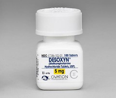

Metamfetamin 5 mg
acute ventricular tachycardia

Image by ADHD CENTER


Orignal From: 236_Team_HeartBit

8/24/2011

Critical Care Ultrasonography (with DVD)

Critical Care Ultrasonography (with DVD)


left ventricular preload - click on the image below for more information.


left ventricular preload


A complete, hands-on guide to successful image acquisition and interpretation at the bedside "The real strength of this textbook is its clinical focus. The editors are to be complimented on keeping a consistent structure within each chapter, beginning with basic physical principles, practical "knobology," scanning tips, key findings, pitfalls and limitations, and how the key findings relate to bedside patho-physiology and decision-making. Thus, the authors have succeeded in providing


Critical Care Ultrasonography (with DVD)


Click on the button for more left ventricular preload information and reviews.


Slide2Call - Stop Accidental Dialing
Slide2Call is a mobile application that prevents you from making unwanted accidental calls. Each attempt to make a call is interrupted by a ready-to-confirm slider. To proceed with the call, simply move the slider from left to right. For Windows Mobile!
Slide2Call - Stop Accidental Dialing
Angiotensin

Article by jekky


Precursor, and types of angiotensin AngiotensinogenAngiotensinogen is an -2-globulin that is produced constitutively and released into the circulation mainly by the liver. It is a member of the serpin family, although it is not known to inhibit other enzymes, unlike most serpins. Plasma angiotensinogen levels are increased by plasma corticosteroid, estrogen, thyroid hormone, and angiotensin II levels.Angiotensinogen is also known as renin substrate.Human angiotensinogen is 452 amino acids long, but other species have angiotensinogen of varying sizes. The first 12 amino acids are the most important for activity.Asp-Arg-Val-Tyr-Ile-His-Pro-Phe-His-Leu-Val-Ile Angiotensin IAsp-Arg-Val-Tyr-Ile-His-Pro-Phe-His-LeuRenin-angiotensin-aldosterone systemAngiotensin I (CAS# 11128-99-7) is formed by the action of renin on angiotensinogen. Renin is produced in the kidneys in response to both decreased intra-renal blood pressure at the juxtaglomerular cells, or decreased delivery of Na+ and Cl- to the macula densa. If more Na+ is sensed, renin release is decreased.Renin cleaves the peptide bond between the leucine (Leu) and valine (Val) residues on angiotensinogen, creating the ten amino acid peptide (des-Asp) angiotensin I (CAS# 9041-90-1).Angiotensin I appears to have no biological activity and exists solely as a precursor to angiotensin 2. Angiotensin IIAsp-Arg-Val-Tyr-Ile-His-Pro-PheAngiotensin I is converted to angiotensin II through removal of two C-terminal residues by the enzyme angiotensin-converting enzyme (ACE, or kinase), which is found predominantly in the capillaries of the lung. ACE is actually found all over the body, but has its highest density in the lung due to the high density of capillary beds there. Angiotensin II acts as an endocrine, autocrine/paracrine, and intracrine hormone.ACE is a target for inactivation by ACE inhibitor drugs, which decrease the rate of angiotensin II production. Angiotensin II increases blood pressure by stimulating the Gq protein in vascular smooth muscle cells (which in turn activates contraction by an IP3-dependent mechanism). ACE inhibitor drugs are major drugs against hypertension.Other cleavage products of ACE, 7 or 9 amino acids long, are also known; they have differential affinity for angiotensin receptors, although their exact role is still unclear. The action of angiotensin II itself is targeted by angiotensin II receptor antagonists, which directly block angiotensin II AT1 receptors.Angiotensin II is degraded to angiotensin III by angiotensinases that are located in red blood cells and the vascular beds of most tissues. It has a half-life in circulation of around 30 seconds, whereas, in tissue, it may be as long as 1530 minutes. Angiotensin IIIAsp | Arg-Val-Tyr-Ile-His-Pro-PheAngiotensin III has 40% of the pressor activity of Angiotensin II, but 100% of the aldosterone-producing activity. Angiotensin IVArg | Val-Tyr-Ile-His-Pro-PheAngiotensin IV is a hexapeptide that, like angiotensin III, has some lesser activity. EffectsSee also Renin-angiotensin_system#EffectsAngiotensins II, III & IV have a number of effects throughout the body: Cardiovascular effectsThey are potent direct vasoconstrictors, constricting arteries and veins and increasing blood pressure.Angiotensin II has prothrombotic potential through adhesion and aggregation of platelets and production of PAI-1 and PAI-2.When cardiac cell growth is stimulated, a local (autocrine-paracrine) renin-angiotensin system is activated in the cardiac myocyte, which stimulates cardiac cell growth through Protein Kinase C. The same system can be activated in smooth muscle cells in conditions of hypertension, atherosclerosis, or endothelial damage. Angiotensin II is the most important Gq stimulator of the heart during hypertrophy, compared to endothelin-1 and A1 adrenoreceptors. Neural effectsAngiotensin III increases thirst sensation (dipsogen) through the subfornical organ (SFO) of the brain, decreases the response of the baroreceptor reflex, and increases the desire for salt. It increases secretion of ADH in the posterior pituitary and secretion of ACTH in the anterior pituitary. It also potentiates the release of norepinephrine by direct action on postganglionic sympathetic fibers. Adrenal effectsAngiotensin II acts on the adrenal cortex, causing it to release aldosterone, a hormone that causes the kidneys to retain sodium and lose potassium. Elevated plasma angiotensin II levels are responsible for the elevated aldosterone levels present during the luteal phase of the menstrual cycle. Renal effectsAngiotensin II has a direct effect on the proximal tubules to increase Na+ reabsorption. It has a complex and variable effect on glomerular filtration and renal blood flow depending on the setting. Increases in systemic blood pressure will maintain renal perfusion pressure, however constriction of the afferent and efferent glomerular arterioles will tend to restrict renal blood flow. The effect on the efferent arteriolar resistance is, however, markedly greater, in part due to its smaller basal diameter; this tends to increase glomerular capillary hydrostatic pressure and maintain glomerular filtration rate. A number of other mechanisms can affect renal blood flow and GFR. High concentrations of Angiotensin II can constrict the glomerular mesangium reducing the area for glomerular filtration. Angiotensin II as a sensitizer to tubuloglomerular feedback preventing an excessive rise in GFR. Angiotensin II causes the local release of prostaglandins, which, in turn, antagonize renal vasoconstriction. The net effect of these competing mechanisms on glomerular filtration will vary with the physiological and pharmacological environment.Renal effects of Angiotensin IITargetActionMechanismRenal artery &afferent arteriolesvasoconstrictionVDCCs Ca2+ influxefferent arteriolevasoconstriction(probably) activate Angiotensin receptor 1 Activation of Gq LC activity P3 and DAG activation of IP3 receptor in SR ntracellular Ca2+mesangial cellscontraction iltration areaactivation of Gq LC activity P3 and DAG activation of IP3 receptor in SR ntracellular Ca2+VDCCs Ca2+ influxTubuloglomerular feedbackIncreased sensitivityIncrease in afferent arteriole responsiveness to signals from macula densamedullary blood flowReduction See alsoACE inhibitorAngiotensin receptorAngiotensin II receptor antagonist References^ Basso N, Terragno NA (December 2001). "History about the discovery of the renin-angiotensin system". Hypertension 38 (6): 12469. doi:10.1161/hy1201.101214. PMID 11751697. http://hyper.ahajournals.org/cgi/pmidlookup?view=long&pmid=11751697. ^ NCBI HomePage^ Physiology at MCG 7/7ch09/7ch09p16^ Skurk T, Lee YM, Hauner H (May 2001). "Angiotensin II and its metabolites stimulate PAI-1 protein release from human adipocytes in primary culture". Hypertension 37 (5): 133640. PMID 11358950. http://hyper.ahajournals.org/cgi/pmidlookup?view=long&pmid=11358950. ^ Gesualdo L, Ranieri E, Monno R, et al. (August 1999). "Angiotensin IV stimulates plasminogen activator inhibitor-1 expression in proximal tubular epithelial cells". Kidney Int. 56 (2): 46170. doi:10.1046/j.1523-1755.1999.00578.x. PMID 10432384. ^ Unless else specified in table, then ref is: Walter F., PhD. Boron (2005). Medical Physiology: A Cellular And Molecular Approaoch. Elsevier/Saunders. ISBN 1-4160-2328-3.  Page 771 Further readingde Gasparo M, Catt KJ, Inagami T, "et al." (2000). "International union of pharmacology. XXIII. The angiotensin II receptors". Parmacol Rev. 52: 415472. PMID 10977869. Brenner & Rector's The Kidney, 7th ed., Saunders, 2004.Mosby's Medical Dictionary, 3rd Ed., CV Mosby Company, 1990.Review of Medical Physiology, 20th Ed., William F. Ganong, McGraw-Hill, 2001.Clinical Physiology of Acid-Base and Electrolyte Disorders, 5th ed., Burton David Rose & Theodore W. Post McGraw-Hill, 2001Lees KR, MacFadyen RJ, Doig JK, Reid JL (1993). "Role of angiotensin in the extravascular system". Journal of human hypertension 7 Suppl 2: S712. PMID 8230088. Weir MR, Dzau VJ (2000). "The renin-angiotensin-aldosterone system: a specific target for hypertension management". Am. J. Hypertens. 12 (12 Pt 3): 205S213S. doi:10.1016/S0895-7061(99)00103-X. PMID 10619573. Berry C, Touyz R, Dominiczak AF, et al. (2002). "Angiotensin receptors: signaling, vascular pathophysiology, and interactions with ceramide". Am. J. Physiol. Heart Circ. Physiol. 281 (6): H233765. PMID 11709400. Sernia C (2002). "A critical appraisal of the intrinsic pancreatic angiotensin-generating system". JOP 2 (1): 505. PMID 11862023. Varagic J, Frohlich ED (2003). "Local cardiac renin-angiotensin system: hypertension and cardiac failure". J. Mol. Cell. Cardiol. 34 (11): 143542. doi:10.1006/jmcc.2002.2075. PMID 12431442. Wolf G (2006). "Role of reactive oxygen species in angiotensin II-mediated renal growth, differentiation, and apoptosis". Antioxid. Redox Signal. 7 (9-10): 133745. doi:10.1089/ars.2005.7.1337. PMID 16115039. Cazaubon S, Deshayes F, Couraud PO, Nahmias C (2006). "[Endothelin-1, angiotensin II and cancer]". Med Sci (Paris) 22 (4): 41622. PMID 16597412. Ariza AC, Bobadilla NA, Halhali A (2007). "[Endothelin 1 and angiotensin II in preeeclampsia]". Rev. Invest. Clin. 59 (1): 4856. PMID 17569300. ... External linksMeSH Angiotensinsv  d  eCardiovascular system, physiology: cardiovascular physiologyHeartVolumesStroke volume = End-diastolic volume End-systolic volumeCardiac output = Heart rate Stroke volumeAfterload  PreloadFrank-Starling law of the heart  Cardiac function curve  Venous return curveAortic valve area calculation  Ejection fraction  Cardiac indexInteraction diagramsCardiac cycle  Wiggers diagram  Pressure volume diagramTropismChronotropic (Heart rate)  Dromotropic (Conduction velocity)  Inotropic (Contractility)  Batmotropic (Excitability)  Lusitropic (Relaxation)Conduction system /Cardiac electrophysiologyCardiac action potential (Atrial action potential, Ventricular action potential)  Effective refractory period  Pacemaker potential  EKG (P wave, PR interval, QRS complex, QT interval, ST segment, T wave, U wave)  Hexaxial reference systemChamber pressureCentral venous pressure/right atrial pressure Right ventricular pressure Pulmonary artery pressure Pulmonary wedge pressure/left atrial pressure Left ventricular pressure Aortic pressureOtherVentricular remodelingVascular system/HemodynamicsBlood flowCompliance  Vascular resistance (Total peripheral resistance)  Pulse  PerfusionBlood pressurePulse pressure (Systolic - Diastolic)  Mean arterial pressureJugular venous pressurePortal venous pressureRegulation of BPBaroreflex  Kinin-kallikrein system  Renin-angiotensin system  Vasoconstrictors/Vasodilators  Autoregulation (Myogenic mechanism, Tubuloglomerular feedback)  Paraganglia (Aortic body, Carotid body, Glomus cell)heart navs: anat/physio/dev, noncongen/congen/neoplasia, symptoms+signs/eponymous, procvascular navs: anat/physio/dev, noncongen/systemic vasculitis/congen/neoplasia, symptoms+signs/eponymous, procv  d  ePeptides: neuropeptidesHypothalamicSomatostatin  CRH  GnRH  GHRH  Orexins  TRH  POMC (ACTH  MSH  Lipotropin)Gastrointestinal hormonesCholecystokinin  Gastric inhibitory polypeptide  Gastrin  Motilin  Secretin  Vasoactive intestinal peptideOther hormonesCalcitonin  Oxytocin  VasopressinOther neuropeptidesAngiotensin  Bombesin  Calcitonin gene-related peptide  Carnosine  Cocaine and amphetamine regulated transcript  Delta sleep-inducing peptide  FMRFamide  Galanin  Galanin-like peptide  Gastrin releasing peptide  Kinins (Bradykinin  Tachykinins)  Neuropeptide S  Neuropeptide Y  Neurophysins  Neurotensin  Pancreatic polypeptide  Pituitary adenylate cyclase activating peptide  VGFNeuromedinsB  N  S  UOpioid peptidesDynorphin  Endomorphin  Endorphin  Enkephalin  Nociceptin  Opiorphinv  d  eAutacoidsKininsKininogen (HMWK, LMWK)  Bradykinin  Kallidin  Tachykinins  Urotensin-IIOthersAngiotensin  Eicosanoid  Histamine  Platelet-activating factor  Serotonin Categories: Human proteins | Peptide hormones | Cardiovascular system | Endocrinology | Physiology


About the Author

I am an expert from Components Electronic suppliers, usually analyzes all kind of industries situation, such as airsoft thompson, skin care private label.


Orignal From: Critical Care Ultrasonography (with DVD)

During isovolumetric ventricular contraction which do the following do: increase, decrease, or do not change?


During isovolumetric ventricular contraction which do the following do: increase, decrease, or do not change?
There are 3 phases in the cardiac cycle:
the cardiac cycle: 1) Ventricular filling: mid-to-late diastole; 2) Ventricular systole; and 3) Isovolumetric relaxation: early diastole. In phase two, ventricular systole, the atria relax and the ventricles begin contracting. Their walls close in on the blood in their chambers, and ventricular pressure rises closing the atriaventricular (AV) valve. Because, for a split second, the ventricles are completely closed chambers and blood volume in the chambers remain constant, it is called the isovolumetric contraction phase.

Good luck.Have a good one always........

Orignal From: During isovolumetric ventricular contraction which do the following do: increase, decrease, or do not change?

8/23/2011

CPAP reversed left ventricular, atrial remodeling.(CARDIOVASCULAR MEDICINE)(continuous positive airway pressure)(Clinical report): An article from: Family Practice News

CPAP reversed left ventricular, atrial remodeling.(CARDIOVASCULAR MEDICINE)(continuous positive airway pressure)(Clinical report): An article from: Family Practice News


left ventricular pressure - click on the image below for more information.


left ventricular pressure


This digital document is an article from Family Practice News, published by International Medical News Group on July 1, 2010. The length of the article is 492 words. The page length shown above is based on a typical 300-word page. The article is delivered in HTML format and is available immediately after purchase. You can view it with any web browser.

Citation Details
Title: CPAP reversed left ventricular, atrial remodeling.(CARDIOVASCULAR MEDICINE)(continuous positive airway pressure)


CPAP reversed left ventricular, atrial remodeling.(CARDIOVASCULAR MEDICINE)(continuous positive airway pressure)(Clinical report): An article from: Family Practice News


Click on the button for more left ventricular pressure information and reviews.


What Is High Blood Pressure?

Article by Clive Harman


Orignal From: CPAP reversed left ventricular, atrial remodeling.(CARDIOVASCULAR MEDICINE)(continuous positive airway pressure)(Clinical report): An article from: Family Practice News

My Normal Heartbeat

My Normal Heartbeat


I have cardiac arrhythmia, this is a recording of my heartbeat when I am well-controlled on medication. I forgot to add in the slide, it is best heard with headphones. The rate in this one is 70-80 which is my normal resting rate. I think there may be one or two PVC's, but I am not sure, it could be my microphone.
Video Rating: 5 / 5


Sudden Cardiac Arrest - Precautionary measures and alternative treatment


Michael Jackson RIP - he died of Sudden Cardiac Arrest ============================================= Sudden cardiac arrest (SCA) occurs when the lower chambers of the heart suddenly stop beating normally and develop what is called ventricular fibrillation (VF). Ventricular fibrillation is a very fast, chaotic heart rate in the lower chambers of the heart. The instant ventricular fibrillation begins, effective blood pumping stops. Ventricular fibrillation quickly becomes more erratic, resulting in sudden cardiac arrest. If this is not corrected immediately via a shock from an external defibrillator or an implantable cardioverter defibrillator (ICD), death will follow. The defibrillator stops the chaotic electrical activity and restores normal heart rhythm.
Video Rating: 5 / 5


Orignal From: My Normal Heartbeat

A rare case of tako-tsubo cardiomyopathy complicated by a left ventricular thrombus.(Case Report): An article from: Southern Medical Journal

A rare case of tako-tsubo cardiomyopathy complicated by a left ventricular thrombus.(Case Report): An article from: Southern Medical Journal


left ventricular thrombus - click on the image below for more information.


left ventricular thrombus


This digital document is an article from Southern Medical Journal, published by Thomson Gale on January 1, 2006. The length of the article is 1943 words. The page length shown above is based on a typical 300-word page. The article is delivered in HTML format and is available in your Amazon.com Digital Locker immediately after purchase. You can view it with any web browser.

Citation Details
Title: A rare case of tako-tsubo cardiomyopathy complicated by a left ventricular thrombus.(Case


A rare case of tako-tsubo cardiomyopathy complicated by a left ventricular thrombus.(Case Report): An article from: Southern Medical Journal


Click on the button for more left ventricular thrombus information and reviews.


Mass Hysteria! 75% payout!
Why are merchant accounts getting shut down left and right? Are you in danger? Yes! Find out the truth about what is really going on at http://www.MassHysteriaReport.com
Mass Hysteria! 75% payout!
Who Should Have An Echocardiography

Article by Armughan Riaz


An Echocardiography is actually the Ultrasound of Heart, this ultrasound produces sound waves and one can have moving picture of heart. This procedure does not involve any radiations and it gives more detail than X-ray. It is a good diagnostic tool for Valvular heart diseases, evaluating pumping function of heart, i.e ejection fraction, in heart attack patients. It is also a good screening test for certain heart disease. However, there are some situations or diseases that one should have an echocardiography test.

Following diseased patients must have an echocardiography. These are the situations in which an echo may influence the clinical management of a patient.

Assessment of valve function, e.g systolic or diastolic murmursAssessment of left ventricular function, systolic diastolic and regional wall motions, e.g suspected heart failure in a patient with breathlessness, or preoperative assessment.Suspected EndocarditisSuspected MyocarditisCardiac TemponadePericardial Disease (e.g Pericarditis) or pericardial effusion, especially if clinical evidence of temponadeComplications of myocardial Infarction, eg MR VSD or pericardial effusion.Suspicion of intracardiac masses- tumour or thrombusCardiac chamber size e.g Left atrial size in atrial fibrillation (AF), Cardiomegaly in chest X-ray.Assessment of artificial valve function.Arrhythmias, e.g Atrial fibrillation, ventricular techycardia (VT)Assessment of right ventricle and right heart Estimation of intracardiac and vascular pressures, e.g pulmonary artery systolic pressures in lung disease and suspected pulmonary hypertensionTo find out cardiac source of embolism in stroke and transient ischaemic attack patients.Exclusion of left ventricular hypertrophy in hypertentionAssessment of congenital heart diseases.These abnormalities are just few and most common that an echo can reveal. For details you may contact your doctor.

What can I expect during an echocardiogram?Often when you visit a Cardiologist, your doctor suggests you for echocardiography of your heart. It is basically the ultrasound of Heart to diagnose various diseases of heart. With the help of sound waves moving picture of heart can be taken.

Echo is a painless and simple procedure without involvement of any radiations. There are no known hazards or risks associated with echocardiography. When you are going to echo room, you will be asked to lie down on a bed and disrobe from the waist up. Doctor or echocardiographer will place electrode on your chest to record ECG during echocardiography. Small amount of gel is applied on your chest and then a small transducer will be placed near the sternum on your chest. Transducer produces sound waves towards the heart.

Echocardiographer may apply some pressure during echo on your chest with transducer. You may be asked to turn your side to left or right depending upon your technician position. You may be asked to hold your breath to take high quality pictures. Then technician will move transducer to different parts of chest to take picture of heart from different angles. Sometimes a dye may be injected before taking echo, as occasionally lungs ribs body tissue prevents sound waves to reach heart muscles. If your heart beat is too fast, then echo may give false result, so echo is avoided in fast tachycardia. A typical echo is performed in about 30-45 minutes. If you have a lung disease, obesity, restlessness or breathlessness, may result in longer test duration. During test, printed pictures are taken by technician from echo machine and later examined by a cardiologist.

Who should have an Echocardiography? Article written by Dr. Armughan Riaz M.B.B.S Dip Card. To Know more about High Blood Pressureand cardiovascular diseases please visit our site.


About the Author

Dr.Armughan maintaining site High Blood Pressure Symptoms Treatment


Orignal From: A rare case of tako-tsubo cardiomyopathy complicated by a left ventricular thrombus.(Case Report): An article from: Southern Medical Journal

EKG: can you have an atrial fibrillation with a premature ventricular complex (pvc)?


EKG: can you have an atrial fibrillation with a premature ventricular complex (pvc)?
Hi purple 22,
..
Yes the two do tend to go together.AF is caused by disorganized electrical conduction in the atria.

It can be treated with medication to slow and regulate the heart or to revert the heart to a normal rhythm.Alternatively it can be reverted by synchronized cardioversion which sounds scary but is a very common procedure which is totally pain free and you will know nothing about it.

AF can last for short or long periods of time during which you may feel palpitations. If you were able to listen to your own heart and feel your pulse at the same time you would notice a marked difference between the two,

Hope this had helped.

love Mel.XHow certain are you that it was a PVC? Yes it can occurr but frequently in atrial fibrillation it could be aberrant ventricular conduction.Aberrant ventricular conduction occurs when a supraventricular impulse reaches the His-Purkinje system when one of the bundle branches (BB) is in the absolute refractory period resulting in slow or blocked conduction through the BB and delayed repolarization through the ventricular muscles causing a BBB (wide Qrs) on the surface ecg in the absence of bundle branch block (BBB) pathology. A right BBB pattern is more common than left BBB. If there is a change in QRS cycle length it is called Ashmans phenonemen. There is a long cycle followed by a short cycle and the beat with the short cycle has a RBBB morphology which causes alot of diagnostic confusion. There is a sudden lengthening of the QRS cycle the subsequent impulse with a shorter cycle or even normal cycle length may be conducted aberrantly . In short aberrant venricular is an abnormal conduction of a supraventricular impulse resulting in a wide QRS complex that can be missed diagnosed as a PVC, but PVC's can occur in atrial fibrillation, you just need to know how to interpret the ecg correctly, clear as mud??? take care, Donna

Orignal From: EKG: can you have an atrial fibrillation with a premature ventricular complex (pvc)?

8/21/2011

Cloudships over Mt. Shasta~

Cloudships over Mt. Shasta~


You will notice that the darker clouds are moving and the other ventricular clouds are standing still. These clouds stayed like this for more than an hour. This is a common almost daily occurrence here in Mt. Shasta, although this was an entire fleet. It felt like the Silver Fleet of the Ashtar Command. After you watch it once, go back and watch it again, except this time watch the foreground, you will see a woman on a horse riding through the ranch pasture.


Emt-D Prehospital Defibrillation


ventricular clouds - click on the image below for more information.


ventricular clouds


Emt-D Prehospital Defibrillation


Click on the button for more ventricular clouds information and reviews.


Orignal From: Cloudships over Mt. Shasta~

Does this sound like supra ventricular tachycardia or ARVD?


Does this sound like supra ventricular tachycardia or ARVD?
well u have some uncontrollable tachycardia since your childhood and it must be benign as you have it since age of 7. if u have a normal blood pressure during the episode it must be a supraventricular one. ARVD is rather nasty disease and it runs in families. diagnosis is impossible if u do not have ecg during the episode. alternative approach is to go for electrophysiologic study while using arrythmia provocation testing.It could be SVT, but there are other things, also. The 24 hour (or 48, or 72) monitor that you are wearing is a good test. You also need an echo (Ultrasound) while exercising, called a stress echo. And I would ask to see an Electrophysiologist, a heart doctor that specializes in the Electrical impulses. It sounds as if you have a serious, real condition. Good for you for quiting the caffeine, dope and cigs. Good Luck!I had a problem like this too over a period of 7 years - it lasted a second or two at first and I saw the doctor's so many times for it, by the time they got the tachycardia on an ekg- it lasted 3 hours and they were more freaked out than me... turned out I had a thyroid problem - and an inexpensive blood test would have picked up on it... so ask the doc to test you for thyroid function...or other things that cause this type of heart function... insist!!!

Hypokalaemia/long QT/Torsades de pointes
ecg ventricular tachycardia

Image by Popfossa


Orignal From: Does this sound like supra ventricular tachycardia or ARVD?

how we can differentiate coarse ventricular fibrillation from ventricular fibrillation?


how we can differentiate coarse ventricular fibrillation from ventricular fibrillation?
Via an ECG trace.By the amplitude of the fibrillation waves.

BTW, it really doesn't matter. Treatment is the same.

Orignal From: how we can differentiate coarse ventricular fibrillation from ventricular fibrillation?

Meaning of Grade 1 Left Ventricular impairment?


Meaning of Grade 1 Left Ventricular impairment?
Most people on here are not doctors. I tried to run a search on this and found where you had asked it on Craigslist too. If it worries you then you should make the doctor sit down and explain it to you or go to another doctor. If your doctor is not willing to discuss you health with you then that is not a good doctor; you need to know about this issue and then you can have some peace of mind. What worries me is if the doctor cannot explain this to you then maybe the doctor is not really knowing what is happening to you.When your heart pumps, it has four chambers and two phases. The Left Ventricle is the chamber that pumps blood to the rest of your body The right pumps it to your lungs to pick up oxygen. Systole is the actual contraction. Diastole is the relaxation. To the ventricles arternate. Contract, then relax. Contract, then relax. The relaxing part allows the chamber to full with blood again so more blood can be pumped around. So putting this into laymen's terms, the part of the heart that is pumping blood to the body is having a little bit of trouble relaxing. Because it has trouble relaxing, it might have trouble pumping blood to the body. Now stop. this sounds scary, but it's not. This is merely a stare 1 problem. Now, if this was a stage 3, you might be in the hospital. I recommend that you listen to what the doctor says and everything should be OK!
best of luck!your condition is mild, that's what grade 1 means.

basically, your heart has to contract and then relax with each heart beat. the contraction helps the heart pump the blood in the left ventricle to the rest of the body, and the relaxation helps the left ventricle fill up with enough blood for the heart to pump with each beat.

you have a mild problem with your left ventricle relaxing. if it was serious, that would mean that your left ventricle was not filling up with enough blood.

but since your left ventricular filling pressure is normal, then things are ok. that just means that your left ventricle was filling up with the normal amount of blood.

your condition is mild, and so don't worry about it. i bet you're probably an older person, since they tend to have this problem.

Orignal From: Meaning of Grade 1 Left Ventricular impairment?

8/20/2011

HELP WITH HEART ventricular tachycardia?


HELP WITH HEART ventricular tachycardia?
go to a cardiologist. ask for a sonogram of the heart. sounds like you might have mitral valve prolapse (weakened mitral valve). It's very common, and symptoms are usually brought on by stress.you are so young, i don't think you have any heart problem. heart problems start at the age of 40 years and upwards. some time kids have congenital heart disease in congenital heart disease tachycardia is common. if you don't have congenital disease then don't worry be happy!

you will never have 100 bpm all the time. it momentary only.you definitly need to see a cardiologist.my son had his first SVT at the age of 2 years old,was put on lanoxin for a year and that was it until last year.he is 12 now.this time he had an episode @school and as he is athmatic thought he is having an attack,so he had some ventolin which only made it worse as ventolin causes fast heart beating.he was transported to hospital and had radiofrequency ablation.he is ok now.
so please see a doc.lots of things can cause a higher rate in beating so dont guess and be safe

Worsening heart failure: rapid heart rate may increase mortality in asymptomatic left-ventricular dysfunction.(Cardiovascular Medicine): An article from: Internal Medicine News


ventricular heart rate - click on the image below for more information.


ventricular heart rate


This digital document is an article from Internal Medicine News, published by International Medical News Group on March 1, 2004. The length of the article is 459 words. The page length shown above is based on a typical 300-word page. The article is delivered in HTML format and is available in your Amazon.com Digital Locker immediately after purchase. You can view it with any web browser.

Citation Details
Title: Worsening heart failure: rapid heart rate may increase mortality in asympto


Worsening heart failure: rapid heart rate may increase mortality in asymptomatic left-ventricular dysfunction.(Cardiovascular Medicine): An article from: Internal Medicine News


Click on the button for more ventricular heart rate information and reviews.


Annotated Sagittal T1 Midline MRI Scan of Reigh's Brain
ventricular heart rate

Image by Reigh LeBlanc


Orignal From: HELP WITH HEART ventricular tachycardia?

The effects of concentric left ventricular hypertrophy on the myocardium Reviews

The effects of concentric left ventricular hypertrophy on the myocardium


concentric ventricular hypertrophy - click on the image below for more information.


concentric ventricular hypertrophy


The effects of concentric left ventricular hypertrophy on the myocardium


Click on the button for more concentric ventricular hypertrophy information and reviews.


Tools For Improving Concentration.
Powerful Techniques To Test, Concentrate And Command Your Mind.
Tools For Improving Concentration.
Hypertensive Heart Disease

Article by Subramani


Hypertensive heart disease is the response of the heart to the increased demands induced by systemic hypertentsion. Pulmonary hypertension also causes heart disease and is referred to as right sided hypertensive heart disease or corpulmonale.

Systmic (left-sided) Hypertensive heart disease

The minimal criteria for the diagnosis of systemic hypertensive heart disease are the following: (1) left ventricular hypertrophy (usually concentric) in the absence of other cardiovascular pathology that might have induced it and (2) a history or pathologic evidence of hypertension. The Framingham heart study established unequivocally that even mild hypertension (levels only slightly above 140/90) mm Hg) if sufficiently prolonged induces left ventricular hypertrophy. Approximately 25% of the U.S. population suffers from hypertension of at least this degree. In hypertension, hypertrophy of the heart is an adaptive response to pressure overload which can lead to myocardial dysfunction, cardiac dilation, congestive heart failure and sudden death.

Compensated systemic hypertensive heart disease may be asymptomatic and suspected only in the appropriate clinical setting by ECG or echocardiographic indications of left ventricular enlargement. As already emphasized, other causes for such hypertrophy must be excluded. In many causes for such hypertrophy must be excluded. In many patients, systemic hypertensive heart disease comes to attention by the onset of atrial fibrillation (owing to left atrial enlargement) or congestive heart failure with cardiac dilation or both. Depending on the severity of the ypertension its duration the adequacy of therapeutic control and underlying basis, the patient may enjoy normal longevity and die of unrelated causes, may develop progressive IHD owing to the effects of hypertension in potentiating coronary atherosclerosis, may suffer progressive renal damage or cerebrovascular accident, or may experience progressive heart failure or sudden cardiac death. There is substantial evidence that effective control of hypertension can prevent or lead to regression of cardiac hypertrophy and its associated risks.

Pulmonary (Right-sided) Hypertensive heart disease (cor pulmonale)

Cor pulmonale, as pulmonary hypertensive heart disease is frequently called, constitutes right ventricular hypertrophy, dilation, and potentially failure secondary to pulmonary hypertension caused by disorders of the lungs or pulmonary vasculature and is the right sided counterpart of left sided(systemic) hypertensive heart disease. Although quite common, right ventricular thickening and dilation caused either by congenital heart disease or by disease of the left side of the heart and the resultant pulmonary venous hypertension owing to postcapillary obstruction to blood flow are exclude from this definition of cor pulmonale.

Based on the suddenness of development of pulmonary hypertension , cor pulmonale may be acute or chronic Acute cor pulmonale can follow massive pulmonary embolism. Chronic cor pulmonale usually implies right ventricular hypertrophy and dilation) secondary to prolonged pressure overload owing to obstruction of the pulmonary arteries or arterioles or compression obliteration of septal capillaries (e.g., owing to primary pulmonary hypertension or emphysema)


About the Author

For more information about Hypertensive heart disease visit http://www.medicalhealthcenter.net


Orignal From: The effects of concentric left ventricular hypertrophy on the myocardium Reviews

Which is worse to have Sustained or Non-sustained Ventricular Tachycardia?


Which is worse to have Sustained or Non-sustained Ventricular Tachycardia?
Ventricular tachycardia lasting more than 30 seconds is called sustained and that which stops before that is Non-sustained. Of course, non-sustained is better than sustained. The need for ICD depends on other factors which only your doctor can decide.Truly this question is a little beyond the scope of friendly advice. In any case simply from the English language one would imagine that non-sustained would be preferable to sustained.
It sounds like your body is struggling with a number of negative factors so anything to improve your general health would be a step in the right direction.

Ablation before ICD implantation can reduce painful shocks: patients at risk for multiple episodes of ventricular tachycardia most likely to benefit.(implantable ... An article from: Heart Advisor


ventricular tachycardia icd - click on the image below for more information.


ventricular tachycardia icd


This digital document is an article from Heart Advisor, published by Belvoir Media Group, LLC on March 1, 2010. The length of the article is 388 words. The page length shown above is based on a typical 300-word page. The article is delivered in HTML format and is available immediately after purchase. You can view it with any web browser.

Citation Details
Title: Ablation before ICD implantation can reduce painful shocks: patients at risk for multiple episodes of ventricular tachycardia


Ablation before ICD implantation can reduce painful shocks: patients at risk for multiple episodes of ventricular tachycardia most likely to benefit.(implantable ... An article from: Heart Advisor


Click on the button for more ventricular tachycardia icd information and reviews.


Orignal From: Which is worse to have Sustained or Non-sustained Ventricular Tachycardia?

Echocardiogram with double inlet left ventricle and post-operative pulmonary artery banding

Echocardiogram with double inlet left ventricle and post-operative pulmonary artery banding


Echocardiogram with double inlet left ventricle and post-operative pulmonary artery banding
Video Rating: 0 / 5


Correctable causes of left ventricular outsow tract obstruction may not be absolute contraindications for arterial switch operation / Sol ventrikul cikis ... of Cardiology (Anadolu Kardiyoloji Dergisi)


left ventricular artery - click on the image below for more information.


left ventricular artery


This digital document is an article from The Anatolian Journal of Cardiology (Anadolu Kardiyoloji Dergisi), published by Thomson Gale on September 1, 2007. The length of the article is 640 words. The page length shown above is based on a typical 300-word page. The article is delivered in HTML format and is available in your Amazon.com Digital Locker immediately after purchase. You can view it with any web browser.

Citation Details
Title: Correctable causes of left ventricular outsow tr


Correctable causes of left ventricular outsow tract obstruction may not be absolute contraindications for arterial switch operation / Sol ventrikul cikis ... of Cardiology (Anadolu Kardiyoloji Dergisi)


Click on the button for more left ventricular artery information and reviews.


My, what a big left ventricular wall you have
left ventricular artery

Image by LucienTj


Orignal From: Echocardiogram with double inlet left ventricle and post-operative pulmonary artery banding

Venous valves within left ventricular coronary veins

Venous valves within left ventricular coronary veins


From the Springer article: Venous valves within left ventricular coronary veins www.springerlink.com by: Anderson, Sara E.; Quill, Jason L.; Iaizzo, Paul A.; The video is organized by vein: anterior interventricular veins, great cardiac veins, posterior veins of the left ventricle, and posterior interventricular veins. At the beginning of each vein, a figure shows its anatomical location. Still images with each representative valve color highlighted aid visualization in subsequent video footage (MPG 19.00 MB) Journal: Journal of Interventional Cardiac Electrophysiology Vol. 23 Issue 2 DOI: 10.1007/s10840-008-9282-6 Published: 2008-10-18
Video Rating: 0 / 5


Bryce Cormier


Join us Sunday, December 28th on CTV from 6 to 8 am for a morning of heroism and inspiration. Our special broadcast, Believing in Heroes, will take you on a journey behind-the-scenes where youll meet some of our bravest heroes. ********** Bryce Cormier was born in February 2005 with a had a serious heart defect, right atrial isomerism which brought on numerous other congenital heart conditions: total anomalous pulmonary vein drainage (TAPVD), where the hearts veins connect to the wrong side causing irregular blood flow; double outlet right ventricle (DORV) which caused her main heart vessels to be on one side, rather than on either side of her heart; and atrial ventricular septal defect. She was rushed to SickKids just after her birth. Her family was told to take her home - essentially to die - because she wasnt strong enough for surgery. Miraculously, she thrived and spent the next few months at home. On July 19, 2006, Bryce was finally strong enough for the surgery and when doctors came out of the operating room, they believed it was a success. For a few hours, she was doing well but sadly, she took a turn for the worse. In the middle of the night, Bryces tiny heart stopped. After manually pumping her heart, SickKids doctors hooked her up to a heart and lung machine that performed the essential functions that her body could no longer do. That night, she also suffered from a stroke. Thankfully, Bryce was stronger than most doctors had ever seen and within months, she was ...
Video Rating: 4 / 5

K.Dextrocardia.levo-TGA.DORV
ventricular vein

Image by HeartBabyHome


Orignal From: Venous valves within left ventricular coronary veins